24.01.2022.
Author: Ante Žuvela, dr.med.dent.
Advantages of lasers in the treatment of granuloma
A granuloma is an inflammation of the tissue that forms around the root of the tooth, most often as a result of a bacterial infection. Caries is one of the most common causes of granulomas.
The granuloma occurs when a tooth infection spreads from the pulp chamber outside the root of the tooth, causing inflammation of the periodontium around the root of the tooth, which is why we often use the term periapical inflammatory response when talking about granuloma.
A granuloma can be seen on X-ray as an enlightenment in the area of the root of the tooth. In order to make accurate diagnosis of granuloma, the patient's medical history, intraoral examination as well as intraoral tests are also very important. A patient with granuloma may experience severe pain, or may also be asymptomatic, completely unaware of having a granuloma. A CBCT is often used as an additional diagnosis, which gives us a more accurate 3D view of the lesion in the bone.
The treatment of granuloma is carried out in such a way that, if necessary, the tooth is anesthetised, so as to relieve any pain. Then a dry working field is established, all caries are removed and all canals found. Once the canals have been found by the doctor, his task is to break them at the top, and accurately measure and widen them. This not only mechanically removes bacteria, infected dentin and residual pulp tissue, but also creates space for good rinsing with disinfectant and laser entry along the entire length of the root itself. The classic treatment of granuloma consists of the mechanical treatment of the canal with needles and disinfection, most often with sodium hypochlorite. However, when applying lasers in the treatment of granuloma, the effect of disinfection on the root canals is much greater. Although ordinary disinfectants are very effective, the possibility of penetration into the lateral canals at the root is very small compared to that with the laser. While disinfectants can eliminate 80% of microorganisms at a depth of 100 µm, lasers are 99.92% effective. Of course, with greater elimination of bacteria, we are more likely to be successful in the therapy itself.
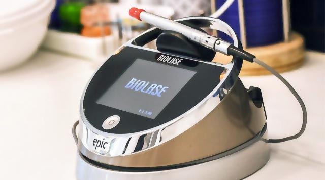
The lasers used in the treatment of granuloma are diode lasers with a wavelength of 840nm and erbium lasers with a wavelength of 2790nm
Once any microorganisms have been successfully eliminated from the canal, they are filled with cement and gutta-percha. As much as the elimination of microorganisms from the canal is important in the treatment, so is a good 3D filling of the canal with filler, as a successful therapy of granuloma.
After the end of treatment, control RVG images are taken immediately after filling, to control the filling itself, and after 3, 6 and 12 months as well. Depending on the size of the granuloma, the bone around the root of the tooth should have been completely restored within a period of 6-12 months after successful endodontic therapy.